Metaphyseal fractures, also known as a bucket handle, chip or corner fracture, occur at the growing end of the bone and only in children. Recent fractures are very difficult to see on x-ray and they are often not associated with any clinical sign of soft tissue swelling or bruising. They may become more obvious radiographically after 11 to 14 days. They are thought to happen when the baby has been pulled or swung violently and the relatively weaker growing point of the bone breaks. They have been noted to occur accidentally following birth injuries, following serial casting of talipes or as a consequence of appropriate physiotherapy to newborn babies. (Source: Core-info leaflet)
The picture of a metaphyseal fracture of an infant’s wrist below comes from a 2000 paper on the orthopaedic aspects of child abuse by Kocher et al and published in the Journal of the American Academy of Orthopaedic Surgeons (http://www.jaaos.org/content/8/1/10.abstract):
A spiral fracture refers to the direction in which the bone is fractured. It implies that there has been a twisting force to cause the fracture. Spiral fractures can also occur accidentally in the femur once the child is walking. (Source: Core-info leaflet)
The picture below of a spiral fracture of the femur in a 2 month old comes from http://www.hawaii.edu/medicine/pediatrics/pemxray/v6c02.html (same website has a wealth of other paediatric radiological images on it if you are interested):
A supracondylar fracture is one in the upper arm immediately above the elbow and is highly suggestive of accidental injury. The picture below comes from http://www.kidsfractures.com/, a site put together by 2 American orthopaedic surgeons who say their aim was to make parents’ experience of having a child with a broken bone a little less traumatic but I think much of the language and many of the pictures are more suited to a medical audience.
A simple linear skull fracture is a break in a cranial bone resembling a thin line, without splintering, depression, or distortion of bone. They are equally prevalent in NAI and in accidental injury. The picture below is of a linear right parietal skull fracture and comes from a Rumanian educational website, http://www.medandlife.ro/medandlife602.html.
This compares with a complex skull fracture which is variously defined as:
• a depressed fracture (where the skull is pushed in)
• two or more fractures of the skull
• fractures that cross the sutures (natural joining edges of skull bones) or those that are widening. (Source: Core-info leaflet)
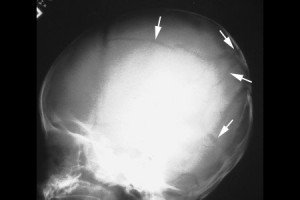
The picture below of a complex skull fracture is from http://www.childabuseconsulting.com/child-abuse-fractures.html which houses other not-very-subtle images of non-accidental burns and bite marks as well.
Rib fractures in infants, particularly posterior ribs, with no history of major trauma are suspicious. The picture below is taken from http://www.learningradiology.com/notes/bonenotes/childabusepage.htm and shows multiple rib fractures with callous formation, the ones of the left 2nd and 6th posterior ribs being the easiest to identify:


